
Illuminant metameric effects on interbrand and intrabrand color differences of direct composite resins
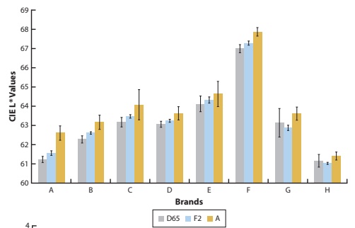
Two restorative materials can appear similar under one light source and different under another. This phenomenon is called illuminant metamerism. This laboratory study examined how different illuminations affected colour differences within and between brands of direct composite resins. It contributes to the study of colour matching beyond a single lighting condition. The journal issue was published in 2022, following online publication in 2021; the available PDF is the advance online version.
Fluorescence intensities of composite resins on photo images
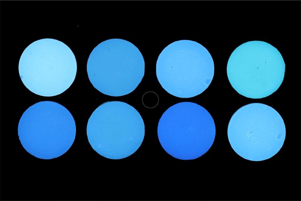
This laboratory study examined whether filtered flash photography could record differences in the fluorescence of composite resins. The researchers tested 64 specimens from eight brands and compared photographic colour measurements with a reference fluorescence measurement. Photographs obtained using 365 nm and 405 nm filters showed a strong relationship with the reference measurements. The work supports photography as a way to document differences between the tested materials; it does not establish the clinical longevity of restorations.
Is the glass half empty or half full? A novel “philosophical” approach to the “mystery” of the so-called ferrule effect
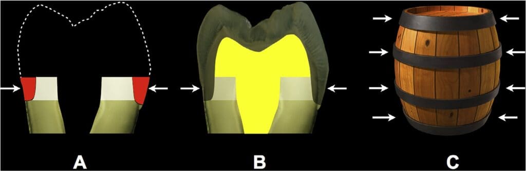
This hypothesis paper revisits the explanation of the so-called ferrule effect in the restoration of endodontically treated teeth. The authors discuss whether the benefit associated with remaining cervical tooth structure can be understood through the resistance form of the preparation and the transfer of lateral forces. The article proposes a framework for further investigation. It is a hypothesis and discussion, rather than a clinical trial demonstrating that a particular restoration is suitable for every tooth.
A Novel Method of Capturing Fluorescence in Clinical Dentistry
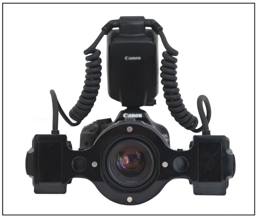
This study describes a photographic setup for recording the fluorescence of teeth and restorations using a macro flash and removable 365 nm filters. The proposed approach was compared with photography using continuous illumination. The authors reported satisfactory fluorescent images under ordinary dental office conditions, with shorter exposure times and without requiring a dark room. The publication focuses on the photographic method and documentation of optical properties.
Digital Photographic Procedure for Comprehensive Two-Dimensional Tooth Shade Analysis
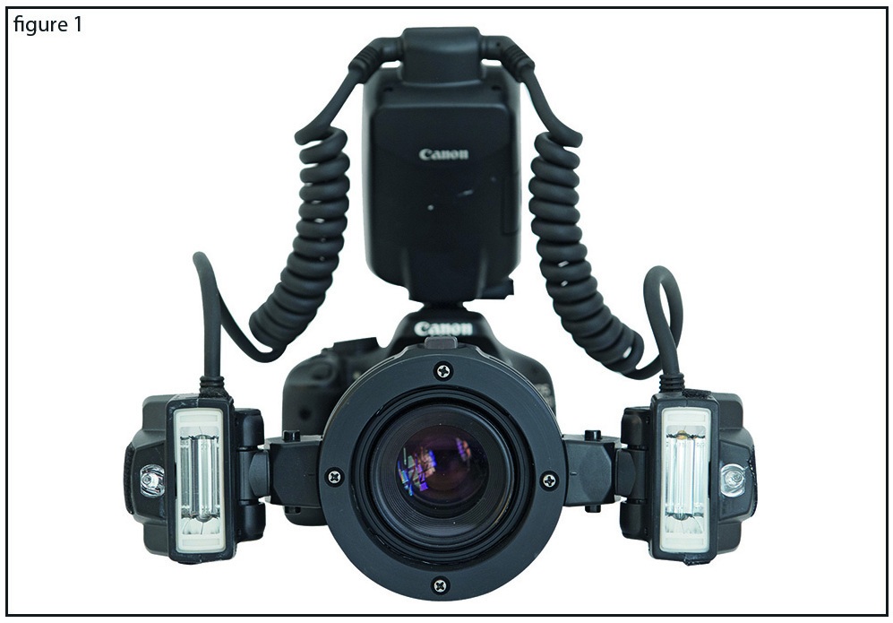
Dental colour matching involves more than selecting a shade tab. This clinical report describes photographic techniques for comparing natural teeth and restorative materials in terms of lightness, fluorescence, opalescence, translucency and metamerism. It presents a DSLR camera setup with additional lighting and filters for documenting these optical properties. The work concerns photographic assessment and communication during restorative treatment.
The i3d Shade Matching Technique: A Case Report
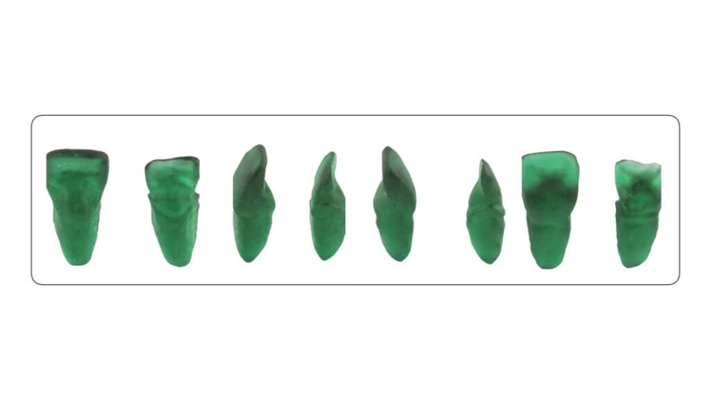
This case report presents the i3d approach to shade matching for direct composite restorations. The technique uses three-dimensional information about tooth anatomy together with customised shade assessment, aiming to reproduce the relationship between enamel and dentin layers. It illustrates a workflow for the reported case. A case report demonstrates an application of a technique but does not establish comparative effectiveness across patients.
Evaluation of enamel thickness of upper anterior teeth in different age groups by dental cone beam computed tomography scan in vivo
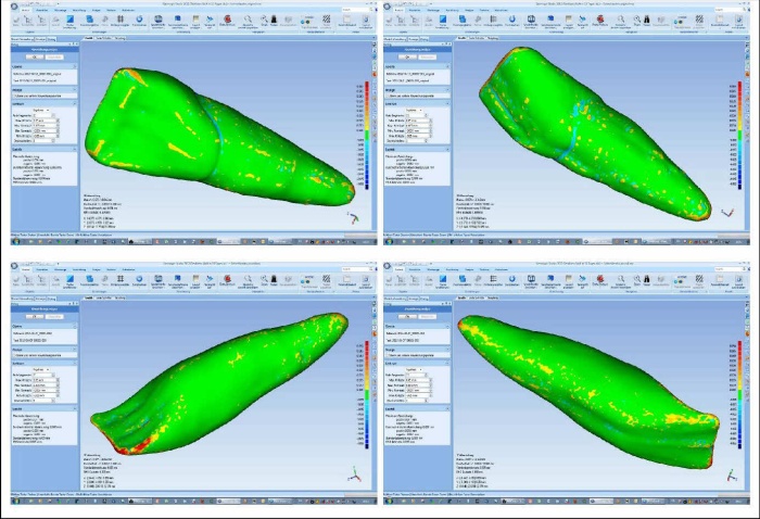
This publication investigates enamel thickness in upper anterior teeth across different age groups using cone beam computed tomography. It addresses the anatomical information relevant to understanding enamel and planning restorative procedures. The illustrated article describes the measurements and analysis in detail. The study is presented here as part of the research archive, with access to the original publication.
The effect of disposable infection control sleeves on the total energy delivered by dental LCUs
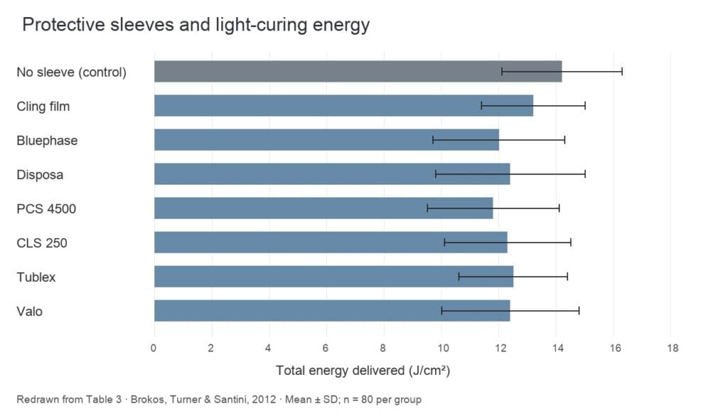
This laboratory study compared seven disposable protective sleeves with four dental light-curing units. Measurements in simulated anterior and posterior tooth positions examined the energy delivered with and without a sleeve. The study found reduced energy delivery when sleeves were used, while the size of the effect also depended on the curing unit and tooth position. The authors discussed testing the complete light-and-sleeve combination. The original publication is available through the bibliographic and full-text links below.
Digital photography in dentistry (2003)
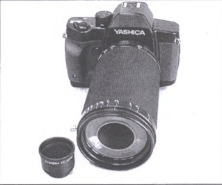
This Greek-language publication discusses dental photography and the transition from conventional to digital photographic equipment. The illustrated article presents cameras, macro lenses, flash systems and examples of dental photographic documentation. Published in 2003, it records the equipment and techniques of that period and forms an early part of the author’s work on photography in dentistry.
 English
 Ελληνικά